Dental X-rays can help your dentist find tooth decay, abscesses, gum disease, and bone loss and prepare for your oral surgery. These X-rays are generally safe but pregnant women should try to avoid X-rays until after the baby is delivered. Digital X-Rays use lower amounts of radiation. A digital X-ray requires less radiation to capture a high-resolution image than the traditional X-rays used a few decades ago. Depending on the type of film, equipment, and image being taken, it may be as much as a 90% reduction in exposure. As such, it’s safe to say that today’s dental X-rays are extremely diagnostic imaging allows dentists to see inside and around the tooth structures where pathology (such as bone loss, oral cancer, or tooth decay) usually located. Diagnosing them as early as possible allows for less-invasive and more cost-effective treatments. Otherwise, such problems can’t be detected until they’ve reached an advanced state that requires more aggressive therapies to manage.
There are two main types of dental x-rays: intraoral (the x-ray film is inside the mouth) and extraoral (the x-ray film is outside the mouth). There are several types of intraoral x-rays which we do with RVG (RadioVisioGraph) . Each shows different aspects of teeth.
Extraoral x-rays are used to detect dental problems in the jaw and skull. There are several types of extraoral x-rays.
 |
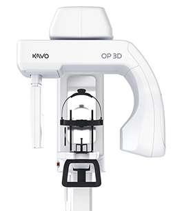
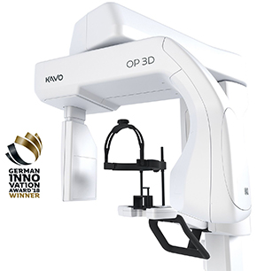
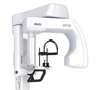
At Keshishian esthetic clinic we use KaVo OP 3D X-ray system. It is a complete X-Ray platform that within 9 seconds provides easy-to-use features throughout the entire dental imaging workflow. With its versatile imaging programs and intuitive user interface, the KAVO OP 3D in its different configurations offers imaging excellence for a variety of users, ranging from general dental practitioners to orthodontists, and all the way to maxillofacial surgeons.